domingo, 22 de marzo de 2015
viernes, 20 de marzo de 2015
L.18 Life in a drop of water
We observed pond water and we found an eucariotic unicelular organism with two flagels.
domingo, 8 de marzo de 2015
L17. Gram staining
MATERIALS
-Hot plate
-1 slide
-1 coverslip
-Tongs
-Needle
-Gram stain (crystal violet, iodine and safranin)
-Ethanol
-Microscope
-Yogurt
PROCEDURE
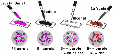
1- First we prepared a heat fixed sample of the bacteria by spreading somre yogurt on a slide and drying it on the hot plate.
2-Then we covered the smear with crystal violet and waited for 1 min. After that we rinsed it with distilled water.
3-We applied iodine solution for another 1 min and again rinsed it with distilled water.
4-Then we decolorized using ethaol. Drop by drop until the purple stops flowing and washed immediately with distilled water.
5-Lastly we covered the sample with safranin stain for and exposure time of 45 seconds and rinsed the sample with tap water.
6-Finally we dried the under part of the slide with paper and viewed it on the microscope.
Results and observations: We saw some bacteria red and other purple. Why?
Gram Positive Cell Wall:
Gram-positive bacteria have a thick
cell wall which is made up of peptidoglycan (50-90% of cell
wall), which stains purple. Peptidoglycan is mainly a polysaccharide
composed of two subunits. The thick
peptidoglycan layer of Gram-positive organisms allows these organisms to
retain the crystal violet-iodine complex and stains the cells as
purple.
Gram Negative Cell Wall:
Gram-negative bacteria have a thinner
layer of peptidoglycan (10% of the cell wall) and lose the crystal
violet-iodine complex during decolorization with the alcohol rinse, but
retain the counter stain Safranin, thus appearing reddish or pink. They
also have an additional outer membrane which contains lipids, which is
separated from the cell wall by means of periplasmic space.
L16. Epidermis cells
On Monday 2nd of March we did two experiments using the microscope, and now i'm going to explain the first one. The objective of this experiment was to identify the shape of epidermis cells, and to identify and explore the part of the stoma and see how it changes its shape when we add salt water.The pores open to facilitate uptake of carbon dioxide
and close to limit the loss of water
MATERIALS
-Slide
-Cover slip
-Tap water
-10% salt water
-Forceps
-Dropeper -Scissors
-Needle
-Leek
PROCEDURE
1-First we cut the stalk of the leek and pulled out the transparent part of the epidermis using forceps.
2-Then we placed the peel into the slide containing a drop of tap water (so the cells don't die) .
3-Next we took a cover slip and placed it gently on the peel with the aid of a needle.
4-We viewed it in the microscope and took pictures of it.
5-Then we prepared a 10% salt solution and put the solution with a dropper on the left part of the slide (so it touched the cover slip) and placed a piece of cellulose paper in the opposite side of the cover slip to let the dissolution go through the sample.
6-Finally we looked through the microscope once more and took more pictures.
Results and observations:
When we first saw the cell we noticed the characteristic shape of a plant cell, a geometric one, a squareand the cell wall. Then we looked closer at it and saw the stomas: they were open. Stoma opens when the guard cells are turgid, when the water potential of the cells adjacent to the guard cells are higher than that in the cell sap of the guard cells and the water molecules from the adjacent cells move into the guard cells by osmosis. The opening of the stoma is an advantage because it allows gaseous exchange to take place.
Then we added salt water and took a second look. Now the stomas were closed because the adjacent cells were hypertonic and the guard cells hypotonic so the water molecules moved out of the guard cells into the adjacent cells by osmosis. When this happens, the guard cells become plasmolysed which in turn causes the stoma to close.
QUESTIONS
1- What is the major function of a cell membrane?
The membrane is selectively permeable to ions and organic molecules and controls the movement of substances in and out of cells. Also it protects the cell from its surroundings.
2- What is the major function of the cell wall?
It surrounds the cell membrane and provides structural support and protection to it. Also it acts as a filtering mechanism and as a pressure vessel, preventing over-expansion when water enters the cell.
3-How does salt affect the cells shapes? And the stomes?
I explained it earlier.
domingo, 1 de marzo de 2015
L13. ANIMAL CELLS vs PLANT CELLS
First we peeled off a leaf from an onion. dyed it with safranin stain (red) and then viewed it in the mycroscope.
Then we did some calculations to know the real size of the cell and its nucleous.
After that, we extracted a cell from our cheeks.
Suscribirse a:
Comentarios (Atom)


